Neurosurgical Innovations and Training Center
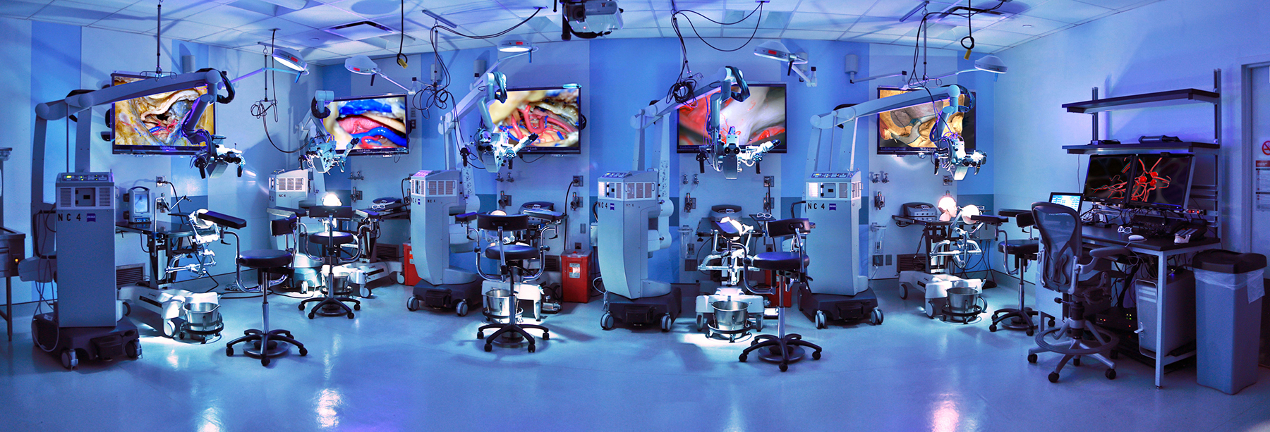
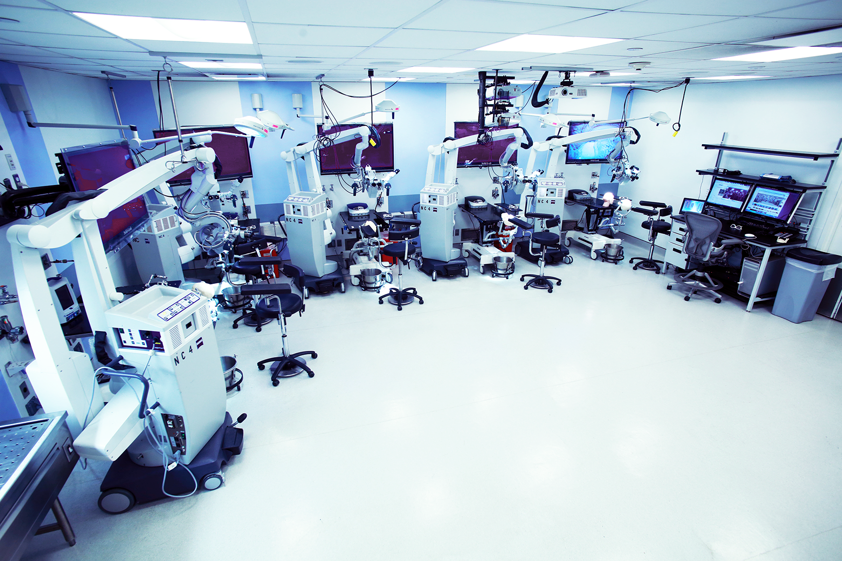
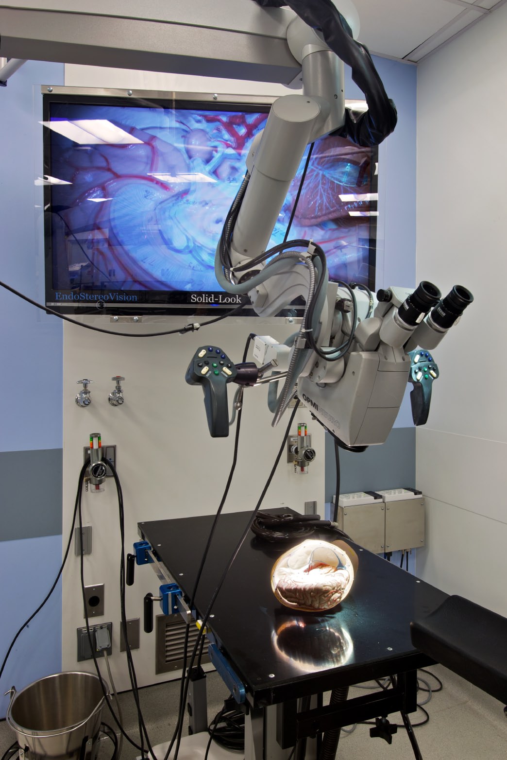
Our 1,000 ft2 dedicated facility contains five adjacent workstations designed to closely recreate the surgical setting of a working operating room. Each workstation is equipped with an adjustable operating table with a Mayfield head holder, a 3D neurosurgical microscope with a xenon light source, a large 3D LCD display, a surgical chair, electric and pneumatic surgical drills, a table mounted adjustable tablet computer, and a complete set of surgical tools.
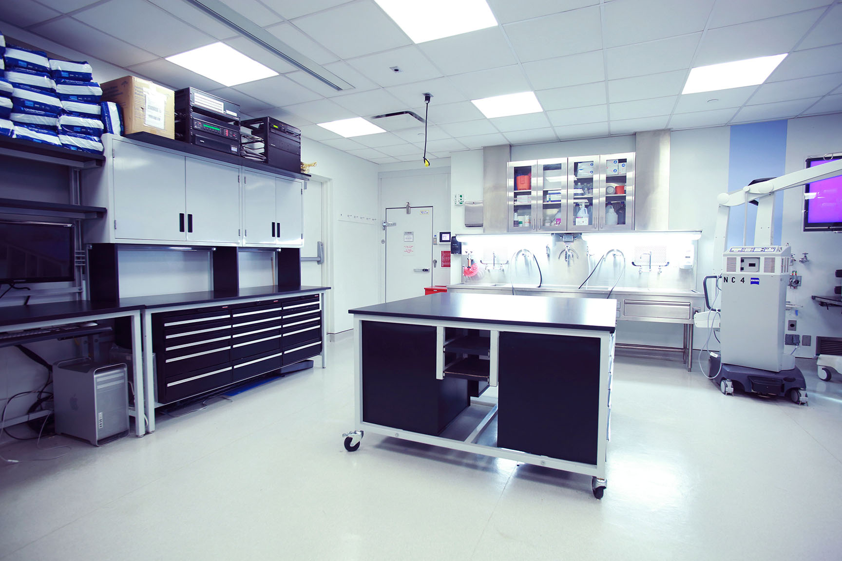
Additionally, each workstation contains surgical lights and wall mounted suction, air, and water lines. A refrigerated cold room within the laboratory is used to store upwards of 200 preserved cadaveric heads, and a large row of sinks with filters capable of handling human tissue is available.
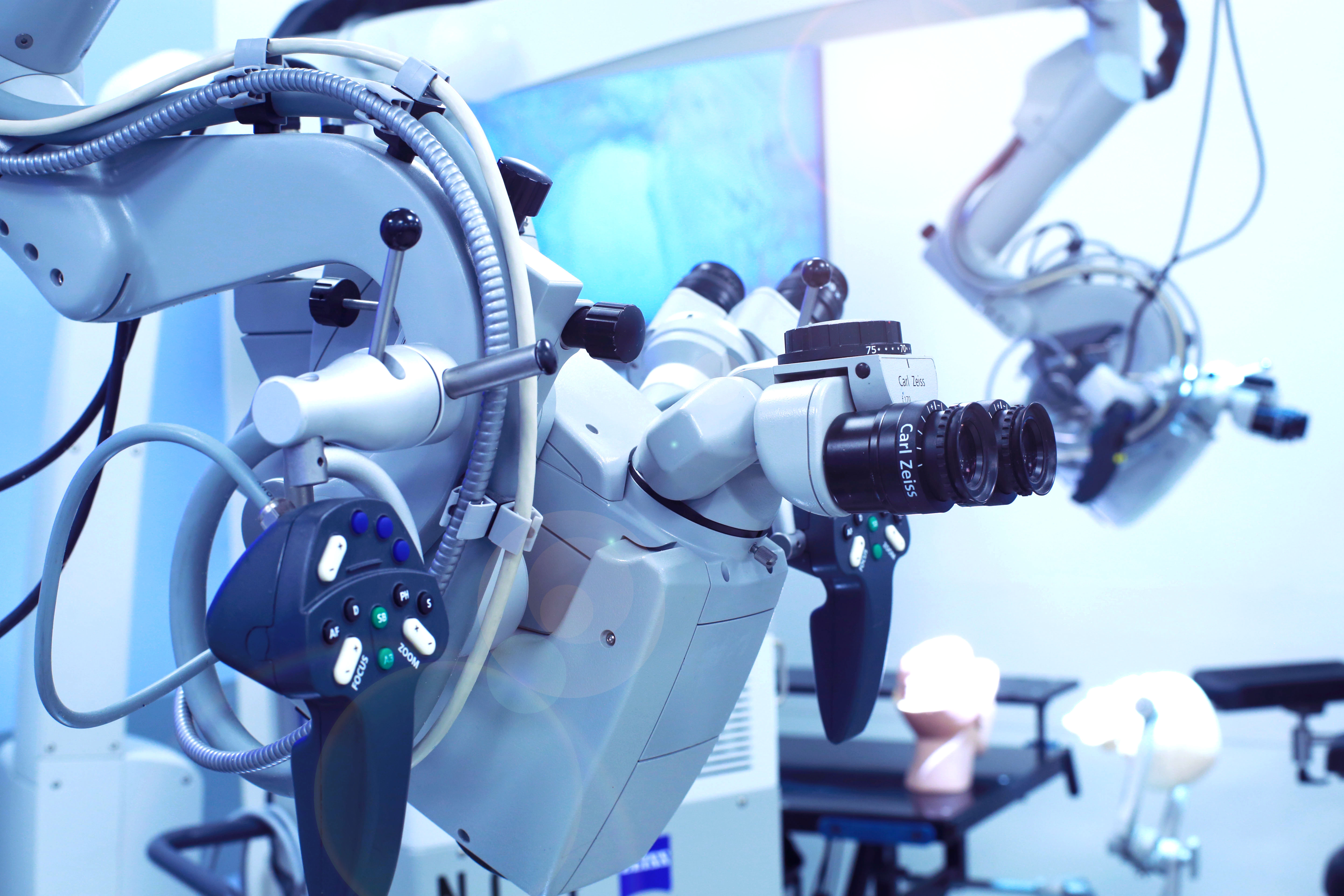
The Zeiss NC-4 neurosurgical microscope at each station is connected to high-definition 3D camera system for screen projection and recording.
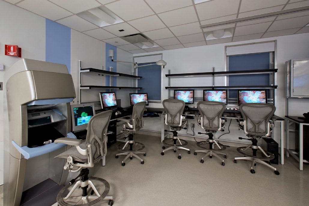
Recordings are obtained and stored using custom designed software. Each camera is capable of optical zoom and can be routed to the 3D display at any workstation, or any of the computer workstations, allowing for simultaneous instruction and dissection. The laboratory is also equipped with a 3D projector with retractable screen that can project the input from any of the lab’s cameras or computers and facilitate didactic lectures.
The lab houses advanced neurosurgical technologies for technologically integrated training, including a neuronavigation system, a surgical robotic simulator, a surgical robot, a Dextroscope holographic surgical planning system, a mobile fluoroscopy system, and virtual reality and computerized simulation for training of surgical procedures. The simulators utilize technology that provides sensory feedback to the user allowing for the simulation of cutting, pulling, and prodding of tissue.
The lab also houses advanced neurosurgical technologies for technologically integrated training.
These technologies include a neuronavigation system, a surgical robotic simulator, a surgical robot, a Dextroscope holographic surgical planning system, a mobile fluoroscopy system, and virtual reality and computerized simulation for training of surgical procedures. The simulators utilize technology that provides sensory feedback to the user allowing for the simulation of cutting, pulling, and prodding of tissue.
We are in the process of building 3D video archives, virtual reality and simulation models, and broadcasting capability for disseminating educational, research, and reference materials to institutions around the world. These initiatives are the first steps in our eventual goal of creating a library of stepwise neurosurgical dissection videos and interactive models and simulations which can be accessed and shared in real-time with surgeons throughout the world via the internet.
- Surgical Table
- Surgical Chair
- Mayfield Head Holder
- Zeiss NC-4 Surgical Microscope
- Electric and Pneumatic Drills
- Surgical Light
- Redundant Suction, Air, and Water Ports
- 3D Microscopic Cameras
- Large 3D LCD Display
- iPad
- Surgical Table and Chair
- Mayfield Head Holder
- Storz 2D Endoscope System
- Visionsense 3D Endoscope System
- Zeiss NC-4 Surgical Microscope
- Electric and Pneumatic Drills
- Tissue Shaver
- Redundant Suction, Air, and Water Ports
- Large 3D LCD Display
- Neuronavigation
- 3D Surgical Planning Systems
- Ultrasonic Bone Cutters
- Synthetic Brain Simulators
- 2D and 3D Projection
- Minimally Invasive Neurosurgery Equipment
- Surgical Simulator
Video Tour